
Traditional microscopy often relies on labeling samples with dyes, but this process is costly and time-consuming. To overcome these limitations, researchers have developed a computational quantitative phase imaging (QPI) method using chromatic aberration and generative AI.
By leveraging the natural variations in focus distances of different wavelengths, the technique constructs through-focus image stacks from a single exposure. With the help of a specially trained diffusion model, this approach enables high-quality imaging of biological specimens, including real-world clinical samples like red blood cells. The breakthrough could revolutionize diagnostics, providing an accessible and efficient alternative to conventional imaging techniques.
Revealing Insights Without Labels
Labeling biological samples with dyes or other agents provides valuable insights, but this method has significant drawbacks that limit its use in clinical diagnostics. It requires time, expensive equipment, and costly reagents. As a result, recent research has focused on label-free microscopy techniques like quantitative phase imaging (QPI).
Unlike traditional imaging methods, QPI analyzes not only the light absorbed or scattered by a sample but also how the sample shifts the phase of light passing through it. This phase shift is directly linked to the sample’s thickness, refractive index, and other structural properties. While QPI typically requires high-end equipment, computational QPI offers a more cost-effective alternative.
The Power of Computational QPI
One of the most widely used computational QPI techniques is based on solving the Transport-of-Intensity Equation (TIE). This mathematical approach reconstructs an image of the sample by analyzing recorded phase changes. It is relatively easy to integrate into existing optical microscopes and produces high-quality images.
However, a major drawback of the TIE method is that it often requires multiple images taken at different focus distances to eliminate artifacts. Collecting these through-focus stacks can be both time-consuming and technically challenging, making TIE-based QPI impractical for many clinical applications.
Making Use of Chromatic Aberration
“Our approach relies on the similar principles as TIE but only needs one image because of a clever combination of physics and generative AI,” says Prof. Artur Yakimovich, Leader of a CASUS Young Investigator Group and corresponding author of the work presented at the AAAI Conference. The information about the phase shift induced by biological specimen does not come from additional exposures taken with other focus distances.
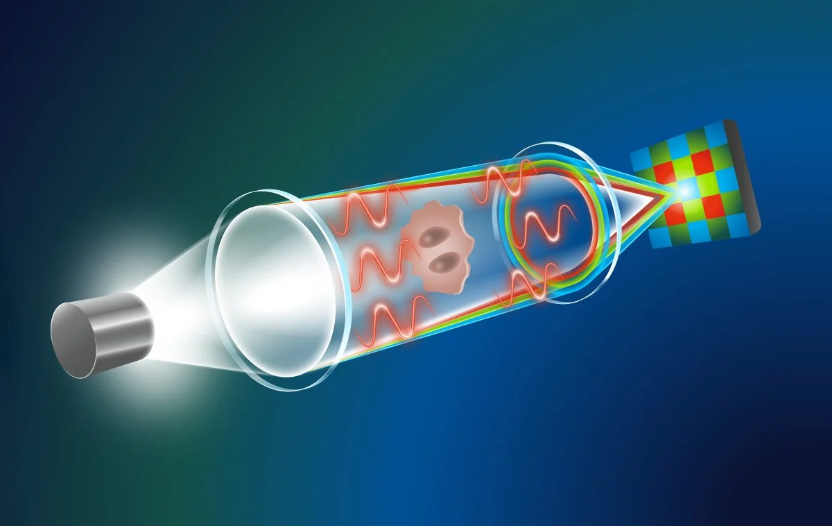
A through-focus stack can also be generated from one single exposure thanks to a phenomenon called chromatic aberration. Most lens systems of the microscope cannot bring all wavelengths of (polychromatic) white light to a single converging point perfectly – a handicap that only highly specialized lenses can correct. This means e.g. red, green and blue (RGB) light have slightly different focus distances.
“By recording the phase shifts of those three wavelengths separately using a conventional RGB detector, one can build a through-focus stack that facilitates computational QPI turning the handicap into an asset,” Yakimovich explains.
AI Overcomes Key Challenges
“Using chromatic aberrations to realize QPI poses one challenge: The distance between the red light focus and the blue light focus is very small,” says Gabriel della Maggiora, PhD student at CASUS and one of the two lead authors of the publication. Solving the TIE the standard way just does not give meaningful results.
“We then reasoned that we could use artificial intelligence. As it turned out, this idea proved to be decisive,” della Maggiora adds. “After training a generative AI model with an open-access data set consisting of 1.2 million images, the model was able to retrieve phase information even though just relying on the very limited data input from the recording.”
Method Validated on Real-World Clinical Specimens
The team drew on a generative AI model for image quality improvement presented last spring: the Conditional Variational Diffusion Model (CVDM). It belongs to a particular family of generative AI models named diffusion models. The developers emphasize that training a CVDM needs significantly less computational effort than training other diffusion models while the results are the same or even better.
Harnessing a CVDM strategy, della Maggiora and colleagues developed a novel diffusion model that is applicable for quantitative data. With this model, they were now able to finally realize computational QPI based on chromatic aberrations.
They validated their generative AI-based approach using for example a common brightfield microscope equipped with a commercially available color camera to make microscopic images from real-world clinical specimen: Analyzing red blood cells in a human urine sample, the method was able to unveil the donut-like shape of these cells whereas another, established computational TIE-based approach was not.
An additional advantage was the virtual absence of cloud artifacts in the images calculated with the new generative AI-based quantitative phase imaging variant.
A New Era for Clinical Microscopy
The Yakimovich group “Machine Learning for Infection and Disease” develops novel computational techniques for microscopy that could be immediately applied in clinical settings. The potential e.g. in diagnostics is huge. Among the techniques used is generative AI. As generative AI is prone to produce hallucinations, a main focus of the group is to reduce them. Incorporating physics-based elements is the key approach here. As the AI-based quantitative phase imaging example shows, this approach is very promising.
Reference: “Single Exposure Quantitative Phase Imaging with a Conventional Microscope using Diffusion Models” by Gabriel della Maggiora, Luis Alberto Croquevielle, Harry Horsley, Thomas Heinis and Artur Yakimovich, 24 December 2024, Proceedings of the AAAI Conference on Artificial Intelligence.